
Ever wondered how our bodies are put together beneath the skin? That’s where a human torso model labeled comes into play—it’s like nature’s blueprint made tangible. These models aren’t just plastic pieces stuck together; they’re an incredibly detailed representation of our anatomy that lets you peek inside the human torso model labeled body without ever picking up a scalpel. Whether you’re a curious learner or a budding doctor, these models bring torso anatomy organs to life.
What is a human torso model labeled?
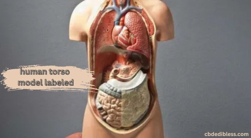
A human torso model labeled is a physical or digital 3D structure that replicates the anatomy of the upper human body. Typically extending from the neck down to the pelvis, it provides a clear, sectioned view of various systems like digestive, respiratory, circulatory, and more. What makes it even more powerful is when it’s labeled—each part is named and often color-coded, making learning far easier. It’s like having a human torso model labeled body textbook turned into a sculpture.
Why Are Labeled Torso Models Important?
If you’ve ever tried memorizing the names of 200+ bones and organs from a flat textbook, you know the struggle is real. Labeled torso models change the game entirely. They make learning anatomy less of a chore and more of a visual adventure. When each organ and part is clearly marked, it removes guesswork and helps students grasp complex body functions with ease. It’s not just about seeing—it’s about understanding and remembering.
Understanding Torso Anatomy
The torso is home to the body’s core systems—literally and functionally. It houses the chest, abdomen, and pelvic regions, where vital organs like the heart, lungs, stomach, liver, and intestines do their work. Understanding torso anatomy is like holding the master key to unlocking the human torso model labeled biology. Whether you’re learning medicine, sports science, or even yoga, knowing what lies beneath the surface improves your understanding tenfold.
External vs Internal View of a Torso Model
On the outside, a torso model might show you the contours of the chest or the muscle structure of the abdomen. But once you remove the outer panels or open up the model, that’s when the real magic happens. Inside, you’ll find a beautifully organized world—lungs nestled beside the heart, intestines curling beneath the liver, kidneys tucked behind it all. The internal view gives a functional map of how everything fits and works together.
Organs Included in a human torso model labeled
Digestive System
This section typically includes the esophagus, stomach, small and large intestines, liver, pancreas, and sometimes the gallbladder. Seeing how food travels and gets broken down inside the body helps demystify digestion.
Respiratory System
You’ll usually find the trachea, lungs, and bronchi clearly labeled. These organs show how we breathe in oxygen and expel carbon dioxide, making the respiratory cycle more understandable.
Urinary System
Models include kidneys, ureters, and bladder, demonstrating how waste is filtered from the blood and expelled from the body.
Reproductive Organs
Depending on the model, you might see either male or female reproductive systems. These typically show organs like the uterus, ovaries, testes, and associated structures in a realistic, labeled layout.
The Role of Muscular and Skeletal Structures
While most focus is on internal organs, many anatomical torso models also highlight key muscular and skeletal components. You’ll often see muscles of the chest wall, rib structures, vertebral column segments, and sometimes even nerve branches. This comprehensive view allows learners to connect the dots between systems—for instance, how breathing affects spinal posture or how muscles protect internal organs.
Anatomy Torso: Detailing Body Systems
Each system in the body doesn’t just exist on its own—it works in harmony with others. An anatomy torso showcases this interconnectedness. For instance, the digestive and circulatory systems are closely tied; the nutrients absorbed in the intestines are sent into the bloodstream. A detailed torso model makes this interplay easier to grasp visually and cognitively.
Anatomical Torso for Medical Students
For med students, an anatomical torso is almost as essential as a stethoscope. These models help students practice identifying organ locations, understanding function, and even prepping for dissections. They act as a bridge between textbook theory and real-life application. And because everything is labeled, it boosts confidence when learning about complex structures in the limited time they have.
How to Read a Labeled Human Torso Model
Start from the top—literally. Begin with the head and neck (if included), then move down to the chest, abdominal cavity, and pelvis. Use the labels as checkpoints. Don’t rush it. Trace each organ, follow the path of the veins or airways, and imagine the body in motion. This interactive process cements knowledge far deeper than rote memorization ever could.
Educational Benefits of Torso Models
Learning is always better when it’s hands-on. A labeled human torso model labeled turns abstract concepts into something you can touch and rotate. It fosters active learning, especially for visual and kinesthetic learners. Whether you’re in a classroom, a hospital training room, or at home prepping for an exam, it allows you to explore the body human torso model labeled in a structured and logical manner.
Choosing the Right Torso Model
Not all models are created equal. Some are basic and only show a few organs. Others are advanced, with cross-sections, removable parts, and detailed labeling. Think about your purpose. Are you just starting to learn, or do you need something for clinical reference? Also, consider size, quality of materials, and whether it comes with a guidebook. Your perfect anatomical companion is out there!
Digital vs Physical Torso Anatomy Models
Technology’s catching up fast. These days, you can use apps or VR to explore torso anatomy organs without ever needing a physical model. But don’t write off the real thing just yet—there’s something uniquely powerful about holding a model in your hands, rotating it, and seeing organs up close. Ideally, combine both for a hybrid learning experience.
Proper Care and Storage of Anatomical Torsos
If you invest in a torso model, take care of it. Keep it out of direct sunlight to avoid fading. Wipe it clean regularly, especially if many hands touch it. Store it in a cool, dry place, preferably in its original packaging. Treat it with respect—it’s your ticket to mastering human anatomy.
Conclusion
A human torso model labeled is more than just a classroom prop—it’s a visual storyteller of the human body. From its perfectly labeled organs to its finely crafted structures, it turns anatomy into an exciting journey of discovery. Whether you’re a student, teacher, or enthusiast, having an anatomical torso at your fingertips makes learning feel personal, human torso model labeled , interactive, and fun. It’s the closest you can get to exploring the wonders of the body without actually opening one up.