
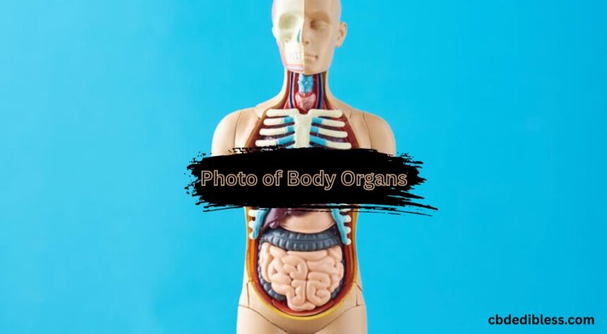
When you think of Photo of Body Organs, you probably imagine stunning landscapes or beautiful portraits. But inside the world of healthcare, photography takes on a whole new life—literally. Photo of body organs play a crucial role in medicine, education, research, and even public awareness. These images give us an intimate view of the structures that keep us alive and functioning, helping doctors diagnose issues, teach students, and allow researchers to make life-saving breakthroughs. It’s like opening a window into a living machine, where every gear and lever has its role—and capturing that in a picture is nothing short of amazing.
The Rise of Medical Imaging in Modern Healthcare
The concept of capturing images of internal body structures isn’t new, but it has drastically transformed over the years. Medical imaging has become the beating heart of diagnostic medicine. From simple black-and-white X-rays to advanced 3D MRIs, the journey has been nothing short of revolutionary. These technologies give us a clear visual roadmap of what’s happening under the skin, often catching problems before they become life-threatening.
From Sketches to Scans: A Brief History
In the early days, anatomy was studied through dissections and carefully hand-drawn illustrations. Artists like Leonardo da Vinci meticulously sketched the human form, creating works that were as beautiful as they were informative. Fast forward to the discovery of X-rays in 1895, and suddenly we could look inside the body without making a single cut. That changed everything.
Evolution of Imaging Technologies
We’ve come a long way since those first blurry X-rays. Now we have CT scans that can create cross-sectional images, MRIs that use magnetic fields to reveal soft tissues in crisp detail, and ultrasounds that use sound waves to show live organ movement. These aren’t just pictures—they’re powerful tools that help decode the mysteries of the human body.
Major Body Organs and Their Photographic Representation
Each organ has its unique structure, function, and Photo of Body Organs. Let’s take a closer look at how different technologies capture these vital parts of us.
The Human Brain: Complex and Captivating
The brain is the command center of the human body. Imaging it requires precision and high-resolution tools because it’s not just about structure—it’s about activity, too. Magnetic Resonance Imaging (MRI) is the gold standard for brain imaging. These scans reveal everything from tumors to strokes and even subtle changes caused by mental health conditions. Seeing a brain MRI is like peeking into the universe of thoughts and emotions.
The Human Heart: Symbol of Life
The heart doesn’t just beat—it tells a story with every thump. Medical imaging of the heart shows us blood flow, valve function, and even how the heart changes during a disease. 3D heart scans are incredibly detailed, and when combined with color Doppler ultrasound, doctors can actually see how blood moves through the chambers. It’s like watching a river run inside your chest.
Lungs: The Breath of Life in Pictures
Our lungs are among the most Photo of Body Organs, especially when captured with contrast in imaging. Their vast network of airways and blood vessels creates a web-like beauty that’s both fragile and functional. Whether it’s pneumonia or a suspicious mass, chest X-rays and CT scans give us a first look. CT images, in particular, slice through the body like a loaf of bread, revealing every nook and cranny of the lungs.
Liver and Kidneys: Detox Duo Captured
These two work silently behind the scenes to cleanse and regulate our body systems. And modern imaging makes it easier than ever to detect problems before symptoms even show. Ultrasound helps check for kidney stones or cysts, while MRI is better for liver issues like fatty liver disease or tumors. These images are essential for monitoring organ health quietly and non-invasively.
Digestive System: Pathways Unveiled
From the esophagus to the intestines, the digestive system is a winding journey that’s rarely seen—unless you have a camera involved. Endoscopy allows real-time visuals of the stomach and intestines using a small camera. It’s one of the few imaging methods where you’re literally looking through the eyes of the scope.
Reproductive Organs: Visualizing Human Continuity
The reproductive organs aren’t just vital for fertility—they’re also central to many health concerns. Ultrasound gives us beautiful images of a fetus developing inside the womb and is also used to detect issues like ovarian cysts or uterine fibroids. These are some of the most emotionally impactful images in medicine.
How These Images Help in Diagnosis
Photo of Body Organs aren’t just snapshots—they’re blueprints for doctors. A good image can reveal the smallest tumors, the tiniest tears, or the first signs of disease. It’s like catching a whisper before it becomes a scream. These images also reduce the need for exploratory surgeries, speeding up treatment and recovery time.
Ethical Considerations in Organ Photography
Taking Photo of Body Organs —especially those from live patients—comes with big responsibilities. There are privacy issues, consent forms, and the need to ensure that these images are used ethically. Whether it’s in a textbook or a research paper, permission and purpose matter.
Educational Value of Organ Photos
Medical students and healthcare workers rely heavily on these images for learning. They’re like the visual textbooks of the body, giving insight into how organs should and shouldn’t look. The ability to compare real Photo of Body Organs to theoretical knowledge is a game-changer in training competent professionals.
How Technology is Making Organ Photography More Accessible
Today, even handheld devices can capture organ images. Portable ultrasounds, smartphone adapters, and cloud storage have made it possible for even remote clinics to benefit from organ imaging. It’s like putting a diagnostic lab in your pocket.
Future of Medical Imaging and Organ Visualization
Artificial Intelligence is now being integrated into imaging systems to spot patterns even expert radiologists might miss. Augmented Reality (AR) could soon allow doctors to “see through” the skin in real time during surgery. The future looks incredibly exciting—and precise.
Best Resources to Access Photo of Body Organs
If you’re curious or learning, websites like the NIH, Radiopaedia, and even open medical textbooks provide verified, high-quality images. Just make sure you’re using them from reputable sources, especially if you’re using them for educational or health-related purposes.
Tips for Interpreting Body Organ Photos Safely
Images can be misleading if not interpreted correctly. Always consult a healthcare professional before jumping to conclusions. These Photo of Body Organs are like maps—you need the right guide to understand them.
Conclusion
Photo of Body Organs is far more than just cool-looking scans. It’s a lifesaving, world-changing field that blends science, art, and technology in the most beautiful way. From diagnostics to education, these images hold power, purpose, and potential. Whether you’re a student, a curious mind, or someone dealing with health issues, understanding these photos can open a whole new world inside the human body.